
In my around 18 months of being a trainee I have now reached my next milestone of 100 brains removed! That means I’ve roughly done slightly less than that in eviscerations I would imagine, as sometimes I have been in the post mortem room and assisted others by just ‘doing the heads’ as we call it. I’m really chuffed that I kept this count and that I’ve got there, it feels good to know. Plus I told the trainee Neuro pathologist who’s been working with us and he said he has done six! I said I just need to get working on what the parts are called now because he puts me to shame naming each and every part, and some bits I really have no idea. I thought it might be a good time to explore the parts of the base of the brain that I’ve learnt so far and what they do.
I don’t think I would ever remember all of that, but here’s an example of how much you could learn!
The brain itself sits in the skull within the meninges, the thick layer outside of the brain but inside the skull is the dura mater. Sometimes this is stuck to the inside of the skull and difficult to remove, other times it can be free. I need to work on getting better at leaving it intact when opening the skull as I often saw through it. I think this is a combination of becoming used to the feeling of the saw but also recognising the differing thickness of skulls. I always think some people must have really heavy heads because the skulls are so thick!
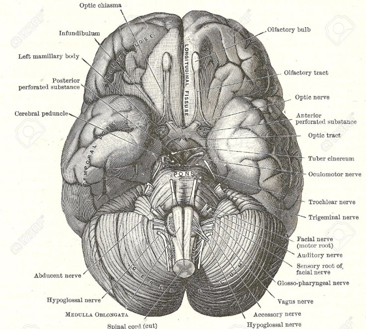
The base of the brain is where the structures that connect to other parts of the body sit. There’s vessels to feed blood to the brain, nerves that shoot off to all the parts of the body too including the spinal cord which is thick and that we disconnect last in order to remove the brain. That’s the bits I know. The only other bit I can name is the rather fancily called Circle of Willis which is a structure of blood vessels at the base of the brain. Sometimes you can identify parts of this that are hard and not working as they should, they are atherosclerotic where there has been a build up in the vessels. It’s taken me a long time to learn how to spell that word, after several times it being spelt letter by letter by the pathologist as I do her notes.

I always think it looks like a little stickman a bit… The Circle of Willis!
The first part of the brain we disconnect as such is the nerves that go to the back of the eyes known as the optic nerves. Once we pull back the front of the brain, just underneath you can see two little white connections that we sever. We work downwards, carefully pulling back the brain and disconnecting the nerves there. The cerebellum, the back of the base of the brain is actually tucked up in a bit more of the dura mater that we have to cut through to remove. Once this is free, we cut down into where the spinal cord has come through, and then can support the top of the brain while scooping out the cerebellum. You then are holding a human brain!
We would, of course, stop at any point if we see anything significant that we might need to note and make the pathologist aware of. This could include any signs of infections, bleeds or other odd things we might not immediately recognise. It takes time to know what is normal and what is not but I’m fairly confident at completing this now.
I hope I’ve explained that fairly well, but if you have any question get in touch, and please bear in mind this is definitely a do not try this at home kind of situation!!
MG x

Ton Up ! Congratulations ! Always (?) wondered how much disconnection was involved in pulling a brain out , never seems that complicated on Silent Witness / CSI etc. 🙂
LikeLiked by 1 person
Thank you very much!
LikeLike